SpeedyCeph Knowledgebase
Technical Support
If you want to learn more, just reach out to us! FYI Technologies will provide telephone support for this product by calling 208-265-8374, Monday - Friday, 8:00 a.m. - 5:00 p.m. PST, excluding holidays. You can also email us at support@fyitek.com.
HIPAA
Keep your Patient Health Information (PHI) secure with SpeedyCeph! We offer enterprise-grade security with end-to-end encryption for all PHI stored by SpeedyCeph. In SpeedyCeph, Patient ID and Last Name are not required. This makes all your PHI de-identified, further increasing the level of security.
Introduction
Welcome to the SpeedyCeph Knowledgebase! SpeedyCeph was designed with the help of AI to be an easy-to-use, yet complex, online cephalometric analysis service. SpeedyCeph is perfect for both orthodontists and general dentists because it combines the advanced features of Dr. Ceph with simple per ceph pricing.
This knowledgebase is meant to describe the general operation of the SpeedyCeph system, and also cephalometrics in general. We will show how variables and constructed landmarks are measured and cover some notable figures in the field of orthodontics. SpeedyCeph is constantly being improved and updated with features from Dr. Ceph. Check out our Development Roadmap!
Getting Started
Purchase a SpeedyCeph analysis on our store to get started tracing cephs. Prices of ceph tracings are based on the complexity of the analysis. Simple analyses, like the ABO, are $20 and complex analyses, like the OSU, are $30. Add your analysis to the cart and go to the Checkout page. At the bottom of the Checkout page is the Patient Details section where you can enter patient information and upload an X-Ray. Within 24 hours of receiving your X-Ray we will trace it for you and send the tracing and analysis results to your email.
Orthodontics
Classes of Malocclusion
Measured from the distal surface of the lower molar to the distal
surface of the upper molar along the occlusal plane.
Class I: The normal position of the upper and lower molars
to each other.
Class II: When the lower molar is seen to lie posterior
(backward or distal) to the upper molar. Also referred to as
distocclusion.
Class II, Division 1: Class II molars and an abnormal
overjet of the incisors. The lower lip is usually displayed under
the upper anterior teeth and the upper lip is sometimes short.
Class II, Division 2: Class II molars with retrusive, or
posteriorly inclined upper anterior teeth. The lower lip is well
formed and both are adequate in length.
Class III: When the lower molar is too far forward of the
upper by at least one cusp. Also referred to as mesiocclusion.
Cephalometrics
Hand tracing every ceph is time consuming, whereas computerized cephalometry is very fast, thus enabling the orthodontist to obtain a more comprehensive diagnostic picture. The need for templates and retracings of acetate overlays is eliminated. An analysis can be performed in a fraction of the time compared to a normal manual registration because it is only necessary to identify the radiological points at the press of a button. The calculations are displayed within seconds. This process removes human error except for errors of landmark identification.


Notable Figures in Orthodontics
Burstone
The Cogs analysis was developed by Dr. Charles J. Burstone when it was presented in 1978 in an issue of AJODO. This was followed by Soft Tissue Cephalometric Analysis for Orthognathic Surgery in 1980 by Arnette et al. In this analysis, Burstone et al. used a plane called horizontal plane, which was a constructed of Frankfurt Horizontal Plane.
Jarabak
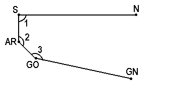
The Jarabak analysis was developed by Dr. Joseph Jaraback in 1972. The analysis interprets how the craniofacial growth may affect the pre and post treatment dentition. The analysis is based on 5 points: Nasion (Na), Sella (S), Menton (Me), Go (Gonion) and Articulare (Ar). They together make a Polygon on a face when connected with lines. These points are used to study the anterior/posterior facial height relationships and predict the growth pattern in the lower half of the face. Three important angles used in his analysis are: 1. Saddle Angle Na, S, Ar 2. Articular Angle S-Ar-Go, 3. Gonial Angle Ar-Go-Me. In a patient who has a clockwise growth pattern, the sum of 3 angles will be higher than 396 degrees. The ratio of posterior height (S-Go) to Anterior Height (N-Me) is 56% to 44%. Therefore, a tendency to open bite will occur and a downward, backward growth of mandible will be observed.
Sassouni
The Sassouni analysis, developed by Dr. Viken Sassouni in 1955,
states that in a well proportioned face, the following four planes
meet at the point O. The point O is located in the posterior cranial
base.This method categorized the vertical and the horizontal
relationship and the interaction between the vertical proportions of
the face.The planes he created are: Palatal Plane (ANS-PNS) Occlusal
Plane (Down's occlusal plane) Mandibular Plane (Go-Me) Plane
parallel inferior border of sella and is parallel to supraorbital
plane Supraorbital plane (Anterior clenoid to roof of orbits) The
more parallel the planes, the greater the tendency for deep bite and
the more non-parallel they are the greater the tendency for open
bite. Using the O as the centre, Sassouni created the following arcs
Anterior Arc - Arc of a circle between the anterior cranial base and
the mandibular plane, with O as the center and O-ANS as the radius.
Posterior Arc - Arc of a circle between anterior cranial base and
mandibular base with O as centre and OSp as radius. Basal Arc - From
A point should pass through B point Midfacial Arc - From Te and
should pass tangent to the mesial surface of the maxillary first
molar.
Center 'O' - a landmark that is the center of the area of
convergence of four planes.
Plane 1: (Parallel Plane)
Posterior Landmark: Clinoidale, (81)
Anterior Landmark: Roof of Orbit, (82)
Reference Landmark: Floor of Sella, (80)
Plane 2: (Palatal Plane)
Posterior Landmark: Posterior Nasal Spine, (45)
Anterior Landmark: Anterior Nasal Spine, (10)
Plane 3: (Occlusal Plane)
Posterior Landmark: Upper Molar Distal Cusp Tip, (23)
Anterior Landmark: Premolar Mesial Contact Point, (46)
Plane 4: (Mandibular Plane)
Posterior Landmark: Gonion, (27)
Anterior Landmark: Menton, (1)


Steiner
Dr. Cecil C Steiner developed the Steiner Analysis in 1953. He used S-N plane as his reference line in comparison to FH plane due to difficulty in identifying the orbitale and porion. Some of the drawbacks of Steiner's analysis includes its reliability on the point nasion. Nasion as a point is known not to be stable due to its growth early in life. Therefore, a posteriorly positioned nasion will increase ANB and more anterior positioned nasion can decrease ANB. In addition, short S-N plane or stepper S-N plane can also lead to greater numbers of SNA, SNB and ANB which may not reflex the true position of the jaws compare to the cranial base. In addition, clockwise rotation of both jaws can increase ANB and counter-clockwise rotation of jaws can decrease ANB.
Tweed
Dr. Charles H. Tweed developed his analysis in 1966. In this analysis, he tried describing the lower incisor position in relation to the basal bone and the face. This is described by 3 planes. He used Frankfurt Horizontal plane as a reference line.
Landmarks
Constructed Landmarks
Geometric Center of Landmarks (Relative to Frankfort
Horizontal) - a landmark that is at the geometric center of four
identifiable landmarks. As used in the Ricketts Analysis to identify
the geometric center of the ramus of the mandible (Xi).
Xi, (500)
Posterior Landmark: Posterior Border of the Ramus, (51)
Anterior Landmark: Anterior Border of the Ramus, (52)
Superior Landmark: Inferior aspect of sigmoid notch, (71)
Inferior Landmark: Inferior to center of sigmoid notch, (72)

Intersection of an Arc and a Plane - a landmark that is the
intersection of an arc and a plane. As used in the Sassouni Analysis
to locate the starting point for the Posterior Arc.
Post Arc & Parallel Plane, (510)
Arc:
Center Landmark: Center 'O', (508)
Reference Landmark: Dorsum Sella, (79)
Modified Plane:
Posterior Landmark: Clinoidale, (81)
Anterior Landmark: Roof of Orbit, (82)
Reference Landmark: Floor of Sella, (80)

Perpendicular Projection to a Plane - a landmark that is
projected onto a plane at the perpendicular. As used in the
Quadrilateral Analysis to identify the posterior end of the mandible
in determining the Mandibular Base Length.
J to GoGn, (507)
Landmark to Project: Point J, (78)
Plane:
Landmark Point 1: Gonion, (27)
Landmark Point 2: Gnathion, (2)

Perpendicular Projection to a Plane using a reference Plane -
a landmark that is projected onto a plane perpendicular to another
plane. As used in the Sassouni Analysis.
Cribiform Perpendicular, (509)
Landmark to Project: Cribiform Point, (85)
Plane 1:
Posterior Landmark: Clinoidale, (81)
Anterior Landmark: Roof of Orbit, (82)
Plane 2:
Posterior Landmark: Floor of Sella, (80)
Anterior Landmark: Floor of Orbit, (84)
Plane to Project to:
Posterior Landmark: Posterior Nasal Spine, (45)
Anterior Landmark: Anterior Nasal Spine, (10)

Intersection of two Planes - a landmark that is the
intersection of two planes. As used in any type of Class III
Prediction Analysis to locate Gonial Intersection.
Gonial Intersection, const, (519)
Plane 1:
Point 1: Articulare, posterior, (31)
Point 2: TangRamus, (142)
Plane 2:
Point 1: Menton, (1)
Point 2: TangGo, (143)

Variables
Two Point Linear Measurement - the linear distance in
millimeters between any two landmarks. Point 1 and Point 2.
e.g. #1 SN
Point 1 = Sella, (35)
Point 2 = Nasion, (38)

Three Point Linear Measurement - the shortest linear distance
in millimeters that Point 3 is from a line connecting Point 1 and
Point 2.
e.g. #22 Upper 1 to NA
Point 1 = Nasion, (38)
Point 2 = Point A, (9)
Point 3 = Upper incisor, incisal edge, (7)

Four Point Linear Measurement - the linear distance in
millimeters of a line from Point 3, perpendicular to a line
connecting Points 1 and 2, to the vertical intersection of Point 4.
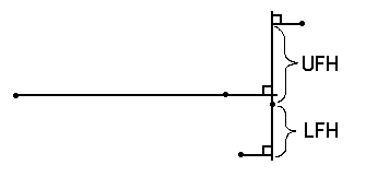
e.g. #82 UFH
Point 1 = Porion, (49)
Point 2 = Orbitale, (40)
Point 3 = Point A, (9)
Point 4 = Nasion, (38)

Three Point Angular Measurement - the inside angle formed by
connecting Point 2 to Point 1 and Point 3 to Point 1. Point 1 being
the convergence of the two lines.
e.g. #9 SNA
Point 1 = Nasion, (38)
Point 2 = Sella, (35)
Point 3 = Point A, (9)

Four Point Angular Measurement - the inside angle formed by a
line connecting Point 1 and Point 2 and a line connecting Point 3
and Point 4. Points 1 and 3 being closest to the convergence of the
two lines.
e.g. #13 Mandibular Plane Angle
Point 1 = Porion, (49)
Point 2 = Orbitale, (40)
Point 3 = Gonion, (27)
Point 4 = Menton, (1)

Distance to Vertical - the shortest linear distance in
millimeters that Point 4 is to a line drawn through Point 3
perpendicular to the line connecting Points 1 and 2.
e.g. #53 WITS
Point 1 = Premolar mesial contact point, (46)
Point 2 = Upper molar distal cusp tip, (23)
Point 3 = Point B, (4)
Point 4 = Point A, (9)

Ratio, Two Linear Measurements - the ratio of one linear
measurement (Point 1) to another linear measurement (Point 2). Point
1 divided by Point 2.
e.g. #84 UFH/LFH
Point 1 = Variable #82 - UFH
Point 2 = Variable #83 - LFH

Angle of Two Planes - the inside angle formed where one
linear measurement (Point 1) converges with a second linear
measurement (Point 2).
e.g. #221 Occlusopalatal Angle
Point 1 = Variable #88 - FOP
Point 2 = Variable #172 - Palatal Plane

Difference, Two Lines - the remainder from subtracting the
linear distance of one linear measurement (Point 2) from the linear
distance of another linear measurement (Point 1).
e.g. #61 Maxillomandibular differential
Point 1 = Variable #11 Effective mandibular length
Point 2 = Variable #10 Effective maxilla length

Sum of Measurements - the result obtained from adding up to
three measurements (Points 1, 2, and 3)
e.g. #81 Sum of Saddle, Articular, Gonial angles
Point 1 = Variable #40 - Saddle angle
Point 2 = Variable #41 - Articular angle
Point 3 = Variable #42 - Gonial angle

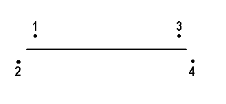
Midpoint Linear Measurement - the linear distance in
millimeters of a line connecting the midpoints of Point 1 and Point
2 and the midpoints of Point 3 and Point 4.

Arc - an arc drawn with Point 1 being the center of the
circle and Point 2 being the reference point (radius). In the Image
View, Angle 1 is the starting point of the arc. If you want the arc
to draw beginning at the reference point (in this example, N), Angle
1 should be 0. If you want the arc to begin drawing 180 degrees from
the reference point, enter 180 in Angle 1. Angle 2 is the ending
point in degrees that the arc will draw.
e.g. - #176 Anterior Arc
Point 1 = Center 'O', (508)
Point 2 = Nasion, (38)

Bisected Angle - the plane created by bisecting the angle
formed by the intersection of two planes. Points 1 and 2 create the
first plane and Points 3 and 4 create the second plane. Points 1 and
3 being closest to the convergence of the two lines.
e.g. #175 Optic Plane
Point 1 = Clinoidale, (81)
Point 2 = Roof of Orbit, (82)
Point 3 = Floor of Sella, (80)
Point 4 = Floor of Orbit, (84)

Modified Plane - a line drawn parallel to another line or
offset by a specified number of degrees. Points 1 and 2 identify the
reference line. Point 3 is the reference point where the new line
will be drawn through. To specify the number of degrees to offset
the parallel line, in the Image View, Arcs and offset, enter the
number of degrees in Angle 1.
e.g. #171 Parallel Plane
Point 1 = Clinoidale, (81)
Point 2 = Roof of Orbit, (82)
Point 3 = Floor of Sella, (80)

Algebraic Formula - the result obtained from solving an
algebraic equation. The formula can contain other variables. For
example, if you want to include variable #283 in the formula, it
would be written as "var283". All three styles of parentheses "{[("
may be used. If a variable is used in the formula, be sure to
include it in the analysis prior to the variable that uses the
Algebraic Formula.
e.g. #285 Max-Mand Differential
Formula = var283 - {90 + [(var284 - 20) * 0.7]}
End-User License Agreement
CAREFULLY READ THE FOLLOWING TERMS AND CONDITIONS. YOUR USE OF THE SOFTWARE WILL INDICATE YOUR ACCEPTANCE TO THE FOLLOWING PROVISIONS. IF YOU DO NOT AGREE WITH THESE TERMS AND CONDITIONS, YOU SHOULD CANCEL YOUR MONTHLY PLAN.
End User License Agreement (EULA) for the SpeedyCeph Monthly
Plan
Introduction: This End User License Agreement ("EULA") is a
legal agreement between you (the "User") and FYI Technologies
("FYI") for the use of the SpeedyCeph web application
("Software"), which includes the web application SpeedyCeph, the
SpeedyCeph Monthly Plan ("Monthly Plan"), and associated media,
printed materials and electronic documentation. By subscribing to
the Monthly Plan, the User agrees to be bound by the terms of this
EULA.
Grant of License: FYI grants the User a non-exclusive,
non-transferable license to use the Software for the purpose of
accessing the web application and any associated services offered
through the Monthly Plan subscription. The User is responsible for
obtaining access to the internet and any additional equipment
necessary to use the Software. The User may use the Software on
any computer, but only for their own individual use. It is not
advised to share login credentials, even with someone that you
know. It is also not advised to use one account for an entire
office.
Restrictions: The User shall not copy, distribute, sell,
resell, or exploit for any commercial purpose the Software or any
portion thereof. The User shall not modify, adapt, translate, or
create derivative works based on the Software or any portion
thereof.
Payment: The User shall pay FYI the Monthly Plan
subscription fee in accordance with the terms of the subscription
and as set forth on FYI's website. The User shall provide FYI with
accurate and complete billing information, including the User's
legal name, address, telephone number, and applicable billing
information. The User shall promptly update this information in
the event of any changes. By agreeing to these terms, the User
agrees to be charged every month for the use of the Software,
until cancelled.
Termination: This EULA shall terminate automatically if the
User breaches any of the terms of this EULA. Upon termination, the
User shall cease all use of the Software and destroy all copies of
the Software in the User's possession. The User may cancel their
membership in the Monthly Plan at any time. Upon cancellation,
your data will be kept for 1 month before being deleted. It is the
User's responsibility to maintain their patient health records
should you decide to cancel the Monthly Plan.
Disclaimer of Warranties: The Software is provided "AS IS"
without warranty of any kind, either express or implied,
including, but not limited to, the implied warranties of
merchantability and fitness for a particular purpose. FYI does not
warrant that the Software will meet the User's requirements or
that the operation of the Software will be uninterrupted or
error-free.
Limitation of Liability: In no event shall FYI be liable
for any damages (including, without limitation, lost profits,
business interruption, or lost information) arising out of the use
or inability to use the Software, even if FYI has been advised of
the possibility of such damages. THIS SOFTWARE MAKES NO DIAGNOSIS
IN REGARD TO THE TREATMENT OF A PATIENT. IT IS INTENDED TO BE ONLY
AN AID TO THE USER IN DETERMINING THE MEASUREMENTS AND VALUES OF A
PATIENT WHEN ESTABLISHING HIS/HER DIAGNOSIS.
Governing Law: This EULA shall be governed by and construed
in accordance with the laws of the United States of America.
HIPAA Law: HIPAA requires that Patient Health Information
("PHI") be secured at all times. FYI employs enterprise-grade
security and AES 256 encryption on all PHI stored by the Software.
If the User's login credentials are kept confidential, only the
User should be able to access their electronic PHI. It is the
User's responsibility to make sure their login credentials are
handled confidentially. As required by HIPAA law, the User may
request copies of confidential PHI stored by FYI or request for
any PHI stored by FYI to be destroyed.
Entire Agreement: This EULA constitutes the entire
agreement between the User and FYI regarding the use of the
Software and supersedes all prior agreements and understandings,
whether written or oral.
By subscribing to the Monthly Plan, the User acknowledges that
the User has read this EULA, understands it, and agrees to be
bound by its terms and conditions.
Bibliography and Further Readings
Ackerman, R.J. (1979) The Michigan school study norms expressed in
template form. Am J Orthod 75:282.
Ackerman, J.L., Proffit, W.R. (1969) The characteristics of
malocclusion: a modern approach to classification and diagnosis.
Am J Orthod 56:443.
Alexander, T.L., Hitchcock, H.P. (1978) Cephalometric standards
for American Negro children. Am J Orthod 74:298-034.
Altemus, L.A. (1960) A comparison of cephalometric relationships.
Angle Orthod 23:217-40.
Altemus, L.A. (1963) Comparative integumental relationships. Angle
Orthod 33:217-21.
Altemus, L.A. (1968) Cephalofacial relationships. Angle Orthod
38:175-89.
Alexander, R.G., Gorman, J.C., Grummons, D.C., Jacobson, R.L.,
Lemchen, M.S. (1990) The DigiGraph work station. Part 2. Clinical
Management. J Clin Orthod 24:403-7.
Anderson, D., Popovich, F. (1989) Correlations among craniofacial
angles and dimensions in Class I and Class II malocclusions. Angle
Orthod 59:37-42.
Anuradha, M., Taneja, J.R., Chopra, S.L., Gupta, A. (1991)
Steiner's norms for North Indian pre-school children. J Ind Soc
Ped Prev Dent 8:36-7.
Argyropoulos, E., Sassouni, V. (1989) Comparison of the
dentofacial patterns for native Greek and American-Caucasian
adolescents. Am J Orthod Dentofacial Orthod 95:238- 49.
Argyropoulos, E., Sassouni, V., Xeniotou, A. (1989) A comparative
cephalometric investigation of the Greek craniofacial patterns
through 4000 years. Angle Orthod 59:195-204.
Ashima, V., John, K.K. (1991) A comparison of the cephalometric
norms of Keralites with various Indian groups using Steiner's and
Tweed's analyses. J Pierre Fauchard Academy 5:17-21.
Athanasiou, A.E., Toutountzakis, N., Mavreas, D., Ritzau, M.,
Wenzel, A. (1991) Alterations of hyoid bone position and
pharyngeal depth and their relationships after surgical correction
of mandibular prognathism. Am J Orthod Dentofacial Orthod
100:259-65.
Athanasiou, A.E., Droschl, H., Bosch, C. (1992) Data and patterns
of transverse dentofacial structure of 6- to 15-year-old children:
A posteroanterior cephalometric study. Am J Orthod Dentofacial
Orthod 101:465-71.
Baughan, B., Demirjian, A., Levesque, G.Y., LaPalme-Chaput, L.
(1979) The pattern of facial growth before and during puberty, as
shown by French-Canadian girls. Ann Hum Biol 6:59-76.
Baumrind, S., Frantz, R.C. (1971) The reliability of head film
measurements. 1. Landmark identification. Am J Orthod 60:111-27.
Baumrind, S. (1980) Computer-aided headfilm analysis: The
University of California San Francisco method. Am J Orthod
78:41-64.
Baumrind, S. (1991) Prediction in the planning and conduct of
orthodontic treatment. In: Melsen B (ed) Current Controversies in
Orthodontics. (Quintessence: Chicago).
Behrents, R.G. (1985) An Atlas of Growth in the Aging Craniofacial
Skeleton. Monograph 17,18, Craniofacial Growth Series. (University
of Michigan, Center for Human Growth and Development: Ann Arbor.)
Bell, W.H., Proffit, W.R., White, R.P. (1980) Surgical Correction
of Dentofacial Deformities, volume I. (WB Saunders:
Philadelphia)137-50.
Ben-Bassat, Y., Dinte, A., Brin, I., Koyoumdjisky-Kaye, E. (1992)
Cephalometric pattern of Jewish East European adolescents with
clinically acceptable occlusion. Am J Orthod Dentofacial Orthop
102:443-8.
Bibby, R.E. (1979) A cephalometric study of sexual dimorphism. Am
J Orthod 76:256-9.
Bishara, S.E., Peterson, J.R., Bishara, E.C. (1984) Changes in
facial dimensions and relationships between the ages of 5 and 25
years. Am J Orthod 85:238-52.
Bishara, S.E., Jakobsen, J.R. (1985) Longitudinal changes in three
normal facial types. Am J Orthod 88:466-502.
Bishara, S.E., Fernandez, A.G. (1985) Cephalometric comparisons of
the dentofacial relationships of two adolescent populations from
Iowa and Northern Mexico. Am J Orthod 88:314-22.
Bishara, S.E., Abdalla, E.M., Hoppens, B.J. (1990) Cephalometric
comparisons of dentofacial parameters between Egyptian and North
American adolescents. Am J Orthod Dentofacial Orthop 97:413-21.
Bolton, W.A. (1962) The clinical application of a tooth-size
analysis. Am J Orhtod 48:504-529.
Bjork, A. (1947) The face in profile. Svenska Tandlak Tid 40 suppl
5B:32-3.
Bjork, A. (1960) The relationship of the jaws to the cranium. In:
Lundstrom A (ed) Introduction to Orthodontics (McGraw-Hill: New
York) 104-40.
Broadbent, B.H. Sr., Broadbent, B.H. Jr., Golden, W.H. (1975)
Bolton Standards of Dentofacial Developmental Growth. (Mosby Year
Book: St Louis.)
Broadbent, B.H. (1983) A new X-ray technique and its application
to Orthodontia. Angle Orthod 1:45-66.
Buck, D.L., Brown, C.M. (1987) A longitudinal study of nose growth
from ages 6 to 18. Ann Plast Surg 18:310-13.
Burstone, C.J. (1958) The integumental profile. Am J Orthod
44:1-25.
Burstone, C.J., James, R.B., Legan, H., Murphy, G.A., Norton, L.
(1978) Cephalometrics for orthognathic surgery. J Oral Surg
36:269-77.
Buschang, P.H., Tanguay, R., Demirjian, A., LaPalme, L.,
Goldstein, H. (1986) Sexual dimorphism in mandibular growth of
French-Canadian children 6 to 10 years of age. Am J Phys Anthropol
71:33-7.
Buschang, P.H., Tanguay, R., Turkewicz, J., Demirjian, A.,
LaPalme, L. (1986) A polynomial approach to craniofacial growth:
description and comparison of adolescent males with normal
occlusion and those with untreated Class III malocclusion. Am J
Orthod Dentofacial Orthop 90:437-42.
Buschang, P.H., Tanguay, R., Demir'ian, A., LaPalme, L.,
Goldstein, H. (1989) Modeling longitudinal mandibular growth:
percentiles for gnathion from 6 to 15 years of age in girls. Am J
Orthod Dentofacial Orthop 95:60-6.
Bushra, A.G. (1948) Variations in human facial pattern in normal
Israelis. Angle Orthod 18:100-2.
Cameron, C. G., Franchi, L., Baccetti, T., McNamara, J. A. (2002)
Long-term effects of rapid maxillary expansion: A posteroanterior
cephalometric evaluation. Am J Orthod Dentofacial Orthod
121:129-35.
Chaconas, S.J., Gonidis, D. (1986) A cephalometric technique for
prosthodontic diagnosis and treatment planning. J Prosthet Dent
56:567.
Chaconas, S.J., Jacobson, R.L., Lemchen, M.S. (1990) The DigiGraph
work station. Part 3. Accuracy of cephalometric analyses. J Clin
Orthod 24:467-71.
Choy, O.W.C. (1969) Cephalometric study of the Hawaiian. Angle
Orthod 39:93-108.
Coben, S.E. (1955) The integration of facial skeletal variants. Am
J Orthod 41:407-34.
Coben, S.E. (1979) Basion Horizontal Coordinate Tracing Film. J
Clin Orthod 13:598- 605.
Coben, S.E. (1986) Basion Horizontal: An Integrated Concept of
Craniofacial Growth and Cephalometric Analysis. (Computer
Cephalometrics Associated: Jenkintown, Pennsylvania.)
Connor, A.M., Moshiri, F. (1985) Orthognathic surgery norms for
American black patients. Am J Orthod 87:119-34.
Costaras, M., Pruzansky, S., Broadbent, B.H. Jr. (1982) Bony
interorbital distance (BIOD), head size and level of the cribiform
plate relative to orbital height: I. Normal standards for age and
sex. J Craniofac Genet Develop Biol 2:19-34.
Cotton, W.N., Takano, W.W., Wong, W.W., Wylie, W.L. (1951) The
Downs' analysis applied to three other ethnic groups. Angle Orthod
21:213-24.
D'Alosio, D., Pangrazio-Kulbersh, V. A comparative and
correlational study of the cranial base in North Americans blacks.
Am J Orthod Dentofacial Orthop 102:449-55.
Davis, D.N., Mackay, F. (1991) Reliability of cephalometric
analysis using manual and interactive computer methods. Br J
Orthod 18:105-9.
Davoody, P.R., Sassouni, V. (1978) Dentofacial pattern differences
between Iranians and American Caucasians. Am J Orthod 73:667-75.
Dawson, P.E. (1989) Evaluation, Diagnosis, and Treatment of
Occlusal Problems. (CV Mosby: St Louis.)
Di Paolo, R.J. (1969) The quadrilateral analysis, cephalometric
analysis of the lower face. J Clin Orthod 3:523-30.
Di Paolo, R.J., Markowitz, J.L., Castaido, D.A. (1970)
Cephalometric diagnosis using the quadrilateral analysis. J Clin
Orthod 4:30-5.
Di Paolo, R.J., Philip, C., Maganzini, A.L., Hirce, J.D. (1983)
The quadrilateral analysis: an individualized skeletal assessment.
Am J Orthod 83:19-32.
Di Paolo, R.J., Philip, C., Maganzini, A.L., Hirce, J.D. (1984)
The quadrilateral analysis: a differential diagnosis for surgical
orthodontics. Am J Orthod 86:470-82.
Di Pietro, G.J., Moergeli, J.R. (1976) Significance of the
Frankfort mandibular plane angle to prosthodontics. J Prosthet
Dent 36:624.
Donatsky, 0., Hillerup, S., Bjorn-Jorgensen, J., Jacobson, P.U.
(1992) Computerized cephalometric orthognathic surgical
simulation, prediction and postoperative evaluation of precision.
Int J Oral Maxillofac Surg 21:199-203.
Downs, W.B. (1948) Variations in facial relationships: their
significance in treatment and prognosis. Am J Orthod 34:812-40.
Downs, W.B. (1952) The role of cephalometrics in orthodontic case
analysis and diagnosis. Am J Orthod 38:162-82.
Downs, W.B. (1956) Analysis of the dentofacial profile. Angle
Orthod 26:191-212.
Droschl, H. (1984) Die fernroentgenwerte unbehandlelter Kinder
zwiscben 6 und 15 Lebensjahr. (Quintessence: Berlin.)
Drummond, R.A. (1968) A determination of cephalometric norms for
the Negro race. Am J Orthod 54:670-82.
DuBrul, E.L. (1980) Sicher's Oral Anatomy. (CV Mosby: St Louis.)
Engel, G.A., Spolter, B.M. (1981) Cephalometric and visual norms
for Japanese population. Am J Orthod 80:48-60.
Enlow, D.H., Moyers, R.E., Hunter, W.S., McNamara, J.A. Jr. (1969)
A procedure for the analysis of intrinsic facial from and growth.
Am J Orthod 56:6-23.
Enlow, D.H., Kuroda, T., Lewin, A.B. (1971) The morphological and
morphogenetic basis for craniofacial form and pattern. Angle
Orthod 41:161-88.
Enlow, D.H., Pfister, C., Richardson, E., Kuroda, T. (1982) An
analysis of Black and Caucasian craniofacial patterns. Angle
Orthod 52:279-87.
Enlow, D.H. (1982) Handbook of Facial Growth, (WB Saunders:
Philadelphia.)
Eriksen, J., Bjorn-Jorgensen, J. (1988) Ortodontisk diagnostik og
behandlings- planlaegning ved hjaelp af digital cefalometri.
Tandlaegebladet 92:499-501.
Farkas, L.G., Sohm, P., Kolar, J,C, Katic, M.J., Munro, I.R.
(1985) Inclinations of the facial profile: art versus reality.
Plast Reconst Surg 75:509-19.
Farkas, L.G., Munro, I.R. (1987) Anthropometric facial proportions
in medicine. Springfield, Il, Charles C. Thomas.
Fischer-Brandies, E., Seeholzer, H., Fischer-Brandies, H., Wimmer,
R. (1991) Die Genauigkeit der Welchteilprofil-Vorhersage mit dem
'Dentofacial Planner' bei skelettaler Progenie. Fortscbr
Kieferorthop 52:289-96.
Fishman, L.S. (1997) Individualized evaluation of facial form. Am
J Orthod Dentofac Orthop 111:510-7.
Flynn, T.R. (1989) Cephalometric norms for orthognathic surgery in
black American adults. J Oral Maxillofac Surg 47:30-8.
Fonseca, R.J., Klein, V.M. (1978) A cephalometric evaluation of
American Negro women. Am J Orthod 73:152-60.
Garcia, C.J. (1975) Cephalometric evaluation of Mexican Americans
using the Downs and Steiner analyses. Am J Orthod 68:67-74.
Genecov, J.S., Sinclair, P.M., Dechow, P.C. (1990) Development of
the nose and soft tissue profile. Angle Orthod 60:191-8.
Gjorup, H., Athanasiou, A.E. (1991) Soft-tissue and dentofacial
profile changes associated with mandibular setback osteotomy. Am J
Orthod Dentofac Orthop 100:312-23.
Graber, T.M. (1972) Orthodontics, Principles and Practice. (WB
Saunders: Philadelphia.)
Grave, K.C., Brown T. (1976) Skeletal ossification and the
adolescent growth spurt. Am J Orthod 69:611-619.
Gravely, J.F., Benzies, P.M. (1991) The clinical significance of
tracing error in cephalometry. Br J Orthod 18: 21-7.
Graven, A.H. (1958) A radiographic cephalometric study of central
Australian aborigines. Angle Orthod 28:12-35.
Grub, J.E. (1992) Computer assisted orthognathic surgical
treatment planning: a case report. Angle Orthod 62:227-34.
Hajighadimi, M., Dougherty, H.L., Garakani, F. (1981)
Cephalometric evaluation of Iranian children and its comparison
with Tweed's and Steiner's standards. Am J Orthod 79:192-7.
Harvold, E.P. (1974) The Activator in Orthodontics. (Mosby Year
Book: St Louis)
Hasund, A., Sivertsen, R. (1969) An Evaluation of Diagnostic
Triangle in Relation to the Facial Type, the Inclination of the
Horizontal Facial Planes and the Degree of Facial Prognathism.
(Acta Universit Bergensis, Medisinske Avhandt: Bergen.)
Hasund, A. (1977) Clinical cephalometry for the Bergen technique.
Orthodontic Department, Dental Institute, University of Bergen:
Bergen.)
Hellsing, E. (1991) Cervical vertebral dimensions in 8-, ll-, and
15-year-old children. Acta Odontol Scand 49:207-13.
Higley, L.B. (1954) Cephalometric standards for children 4 to 8
years of age. Am J Orthod 40:51-9.
Hing, N.R. (1989) The accuracy of computer generated prediction
tracings. Int J Oral Maxillofac Surg 18:148-51.
Hofrath, H. (1931) Die Bedeutung der Roentgenfern und
Abstandsaufnahme für Diagnostik der Kieferanomalien. Fortscbr
Kieferortbop 1:232-48.
Holdaway, R.A. (1983) A soft-tissue cephalometric analysis and its
use in orthodontic treatment planning. Part I, Am J Orthod
84:1-28.
Holdaway, R.A. (1984) A soft-tissue cephalometric analysis and its
use in orthodontic treatment planning. Part II, Am J Orthod
85:279-93.
Houston, W.J.B. (1982) A comparison of the reliability of
measurements of cephalometric radiographs by tracings and direct
digitalization. Swed Dent J suppl 15:99-103.
Humerfelt, A. (1978) A roentgenographic cephalometric
investigation of Norwegian children with normal occlusion. Scand J
Dent Res 78:117-43.
Hunter, W.S., Baumrind, S., Moyers, R.E. (1993) An inventory of
United States and Canadian growth record sets: Preliminary report.
Am J Orthod Dentofacial Orthop 103:545-55.
Iijuka, T., Ishikawa, F. (1957) Normal standards for various
cephalometric analyses in Japanese adults. J Jap Orthod Soc
16:4-12.
Ingerslev, C.H., Solow, B. (1975) Sex differences in craniofacial
morphology. Acta Odont Scand 33:85-94.
Ingervall, B., Schmoker, R. (1990) Effect of surgical reduction of
the tongue on oral stereognosis, oral motor ability, and the rest
position of the tongue and mandible. Am J Orthod Dentofacial
Orthop 97:58-65.
Ioannidou-Marathlotou, J., Kolokithas, G. (1991) Etude des
compensations dans laformation harmonieuse du visage des adultes
Grecs. L'Orthodontie Française 62:811-27.
Isaacson, R.J., Lindauer, S.J., Strauss, R.A. (1991) Computers and
cephalometrics. Alpha Omega 84:37-40.
Jacobson, A. (1975) The "Wits" appraisal of jaw disharmony. Am J
Orthod 67:125-38.
Jacobson, A. (1976) Application of the "Wits" appraisal. Am J
Orthod 70:179-89.
Jacobson, A. (1978) The craniofacial skeletal pattern of the South
African Negro. Am J Orthod 73:681-91.
Jacobson, A. (1979) The proportionate template as a diagnostic
aid. Am J Orthod 75:156-172.
Jacobson, A., Caufield, P.W. (1985) Introduction to Radiographic
Cephalometry. (Lea and Febiger: Philadelphia.)
Jacobson, A. (1990) Planning for orthognathic surgery - art or
science? Int J Adult Orthod Orthognath Surg 5:217-24.
Jarabak, J.R., Fizzell, J.A. (1972) Technique and Treatment with
Lightwire Edgewise Appliance. (CV Mosby: St Louis.)
Jenkins, D.H. (1955) Analysis of orthodontic deformity employing
lateral cephalostatic radiography. Am J Orthod 41:442-52.
Jones, O.G. (1989) A cephalometric study of 32 North American
black patients with anterior open bite. Am J Orthod Dentofacial
Orthop 95:289-96.
Kavadia-Tsatala, S. (1989) The maxilla in harmonious face. A
cephalometric study in adult Greeks (Ricketts' ten factor
analysis). Orthod Review 1:5-22.
Kavadia-Tsatala, S., Topouzelis, N., Sidiropoulou, S., Markovitsi,
H., Kolokithas, G. (1989) Establishment of normal cepohalometric
values of adults with optimal occlusion and harmonious face.
Orthod Review 1:87-104.
Kiyak, H.A., Hohl, T., West R.A. et al (1981) Sex differences in
motives for and outcomes of orthognathic surgery. J Oral Surg
39:757-764.
Kolokithas, G. (1981) Establishment of characteristic values of
skull morphology of Greeks by means of cephalometric radiography.
Thessaloniki: Aristotle University of Thessaloniki.
Korn, E.L., Baumrind, S. (1990) Transverse development of the
human jaws between the age of 8.5 and 15.5 years studied
longitudinally with use of implants. J Dent Res 69:1298-306.
Kowalsky, C.J., Walker, G.F. (1971) Distribution of the mandibular
incisor-mandibular plane angle in normal individuals. J Dent Res
50:984-1690.
Kowalsky, C.J., Nasjleti, C., Walker, G.F. (1974) Differential
diagnosis of American adult male black and white populations using
Steiner's analysis. Angle Orthod 44:346-50.
Krogman, W.M., Sassouni, V. (1957) A syllabus in roentgenographic
cephalometry. (Philadelphia Center for Research in Child Growth:
Philadelphia.)
Lavelle, C. (1974) Craniofacial profile angles in Caucasians and
Negroes. J Dent Res 2:160-6.
Legan, H., Burstone, C.J. (1980) Soft tissue cephalometric
analysis for orthognathic surgery. J Oral Surg 38:744-51.
Lestrel, P.E., Roche, A.F. (1986) Cranial base shape variation
with age: a longitudinal study of shape using Fourier analysis.
Hum Biol 58:527-740.
Lew, K.K. (1992) The reliability of computerized cephalometric
soft tissue prediction following bimaxillary anterior subapical
osteotomy. Int J Adult Orthod Orthognath Surg 7:97-101.
Lew, K.K., Ho, K.K., Keng, S.B., Ho, K.H. (1992) Soft tissue
cephalometric norms in Chinese adults with esthetic facial
profiles. J Oral Maxillofac Surg 50:1184-9.
Lew, K.K., Tay, D.K. (1993) Submentovertex cephalometric norms in
male Chinese subjects. Am J Orthod Dentofacial Orthop 103:247-52.
Lewis, A.B., Roche, A.F., Wagner, B. (1985) Pubertal spurts in
cranial base and mandible: comparisons between individuals. Angle
Orthod 55:17-30.
Lewis, A.B., Roche, A.F. (1988) Late growth changes in the
craniofacial skeleton. Angle Orthod 58:127-35.
Lim, J.Y. (1992) Parameters of Facial Asymmetry and their
Assessment. (Department of Orthodontics and Pediatric Dentistry:
Farmington, Connecticut.)
Liu, Y.T., Gravely, J.F. (1991) The reliability of the Ortho Grid
in cephalometric assessment. Br J Orthod 18:21-7.
Lundstrom, A., Woodside, D.G., Popovich, F. (1989) Panel
assessments of the facial profile related to mandibular growth
direction. Eur J Orthod 11:271-297.
Lundstrom, A., Forsberg, C.M., Peck, S., McWllllam, J. (1992) A
proportional analysis of the soft tissue facial profile in young
adults with normal occlusion. Angle Orthod 62:127-33.
Lundstrom F., Lundstrom, A. (1992) Natural head position as a
basis for cephalometric analysis. Am J Orthod Dentofacial Orthop
101:244-247.
Macri, V., Wenzel, A. (1993) Reliability of landmark recording on
film and digital lateral cephalograms. Eur J Orthod 15:137-48.
Margolis, H.I. (1939) A plastic and graphic technique for
recording dental changes and facial growth. Am J Orthod
25:1027-36.
Mazaheri, M., Krogman, W.M., Harding, R.L., Millard, R.T., Mehra,
S. (1977) Longitudinal analysis of growth of the soft palate and
nasopharynx from six months to six years. Cleft Palate J 14:52-62.
McMinn, R.M.H., Hutchings, R.T. (1977) A Colour Atlas of Human
Anatomy. (Wolfe: London.)
McNamara, J.A. Jr. (1983) A Method of Cephalometric Analysis.
Center for Human Growth and Development, University of Michigan,
Ann Arbor.
McNamara, J.A. Jr. (1984) A method of cephalometric evaluation. Am
J Orthod 86:449-69.
McNamara, J.A. Jr., Brudon, W.L. (1993) Orthodontic and Orthopedic
Treatment in the Mixed Dentition. (Needham Press: Ann Arbor.)
McNamara, J.A. Jr., Brust, E.W., Riolo, M.L. (1993) Soft tissue
evaluation of individuals with an ideal occlusion and a
well-balanced face. In: McNamara JA Jr (ed) Esthetics and the
Treatment of Facial Form. Monograph 28, Craniofacial Growth
Series. (University of Michigan, Center for Human Growth and
Development: Ann Arbor.)
Melsen, B., Athanasiou, A.E. (1987) Soft Tissue Influence in the
Development of Malocclusion. (The Royal Dental College: Aarhus.)
Meng, H., Goorhuis, J., Kapila, S., Nanda, R.S. (1988) Growth
changes in the nasal profile from 7 to 18 years of age. Am J
Orthod Dentofacial Orthop 94:317-26.
Micheils, L.Y., Tourne, L.P. (1990) Nasion true vertical: a
proposed method for testing the clinical validity of cephalometric
measurements applied to a new cephalometric reference line. J
Adult Orhtod Orthog Surg 5:43-52.
Miura, F., Inoue, N., Suzuki, K. (1963) The standards of Steiner's
analysis for Japanese. Bull Tokyo Med Dent Univ 10:387-95.
Moorrees, C.F.A, Lebret, L. (1962) The mesh diagram and
cephalometrics. Angle Orthod 32:214-231.
Moyers, R.E. (1973) Handbook of Orthodontics, ed 3, (Mosby Year
Book: Chicago.)
Moyers, R.E., Bookstein, F.L., Hunter, W.S. (1988) Handbook of
Orthodontics. (Mosby Year Book: Chicago.)
Nanda, R., Nanda, R.S. (1969) Cephalometric study of the
dentofacial complex of North Indians. Angle Orthod 39:22-8.
Nanda, R.S., Meng, H., Kapila, S., Goorhuis, J. (1990) Growth
change in soft tissue facial profile. Angle Orthod 60:177-90.
O'Reilly M, Yanniello GJ, Mandibular growth changes and maturation
of cervical vertebrae. A longitudinal cephalometric study. Angle
Orthod 1988; 58:179-184.
Oliver, R.G. (1991) Cephalometric analysis comparing five
different methods. Br J Orthod 18:277-83.
Park, I.C., Bowman, D., Klapper, L. (1989) A cephalometric study
of Korean adults. Am J Orthod Dentofacial Orthop 96:54-9.
Pedersen, E., Eriksen, J., Gotfredsen, E. (1988) Computerized
Orthodontic Treatment Planning. (Department of Orthodontics,
Aarhus University: Aarhus.)
Phillips, C., Devereux, J.P., Tulloch, J.F.C., Tucker, M.R. (1986)
Full-face soft tissue response to surgical maxillary intrusion.
Int J Adult Orthod Orthognath Surg 1:299-304.
Platou, C., Zachrisson, B.U. (1983) Incisor position in
Scandinavian children with ideal occlusion: a comparison with the
Ricketts and Steiner standards. Am J Orthod 83:341-52.
Popovich, F., Thompson, G.W. (1977) Craniofacial templates for
orthodontic case analysis. Am J Orthod 71:406-20.
Popovich, F., Thompson, G.W. (1988) Craniofacial templates for
orthodontic case analysis. In: Clark's Clinical Dentistry. (JB
Lipincott: Philadelphia.)
Proffit, W.R. (1991) Treatment planning: The search for wisdom.
Surgical Orthodontic Treatment. (Mosby Year Book: St. Louis).
Proffit, W.R., Fields, H.W. (1993) Contemporary Orthodontics.
(Mosby Year Book: St. Louis)
Rakosi, T. (1982) An Atlas and Manual of Cephalometric
Radiography. (Wolfe: London.)
Richardson, A. (1981) A comparison of traditional and computerized
methods of cephalometric analysis. Eur J Orthod 3:15-20.
Richardson, E.R., Malhatru, S.K. (1974) Vertical growth of the
anterior face and cranium in inner city Negro children. Am J Phys
Anthropol 41:361-6.
Richardson, E.R. (1980) Racial differences in dimensional traits
of the human face. Am J Orthod 50:301-311.
Richardson, E.R. (1991) Atlas of Craniofacial Growth in Americans
of African Descent. Monograph 26, Craniofacial Growth Series.
(University of Michigan, Center for Human Growth and Development:
Ann Arbor.)
Ricketts, R.M. (1956) Role of cephalometrics in prosthetic
diagnosis. J Prosthet Dent 6:488.
Ricketts, R.M. (1957) Planning treatment on the basis of the
facial pattern and an estimate of its growth. Am J Orthod
27:14-37.
Ricketts, R.M. (1960) The influence of orthodontic treatment on
facial growth and development. Angle Orthod 30:103-33.
Ricketts, P.M. (1960) A foundation for cephalometric
communication. Am J Orthod 46:330-57.
Ricketts, R.M. (1968) Esthetics, environment and the law of lip
relation. Am J Orthod 54:272-89.
Ricketts, R.M. (1970) The sources of computerized cephalometrics.
In: Ricketts RM, Bench RW (eds) Manual of Advanced Orthodontics
Seminar.
Ricketts, R.M., Bench, R.W., Hilgers, J.J., Schulhof, R. (1972) An
overview of computerized cephalometrics. Am J Orthod 61:1-28.
Ricketts, R.M. (1972) The value of cephalometrics and computerized
technology. Am J Orthod 42:179-99.
Ricketts, R.M. (1975) A four-step method to distinguish
orthodontic changes from normal growth. J Clin Orthod 9:208-28.
Ricketts, R.M., Bench, R.W., Gugino, C.F., Hilgers, J.J.,
Schulhof, R. (1979) Bioprogressive therapy. (Rocky Mountain
Orthodontics: Denver).
Ricketts, R.M. (1981) Perspectives in the clinical application of
cephalometrics. Angle Orthod 51:115-50.
Ricketts, R.M. (1981) The Golden Divider. J Clin Orthod 15:725-59.
Ricketts, R.M. (1991) Orthodontics today - a total perspective.
Mechanical and Biological Basics in Orthodontic Therapy. (Huthig
Buch Verlag: Heidelberg) 249-308.
Riedel, R.R. (1952) The relation of maxillary structures to
cranium in malocclusion and in normal occlusion. Angle Orthod
22:142-5.
Riolo, M.L., Moyers, R.E., McNamara, J.A. Jr., Hunter, W.S. (1979)
An Atlas of Craniofacial Growth: Cephalometric Standards from the
University School Growth Study. Monograph 2, Craniofacial Growth
Series. (University of Michigan, Center for Human Growth and
Development: Ann Arbor.)
Roche, A.F., Mukherjee, D., Guo, S. (1986) Head circumference
growth patterns: birth to 18 years. Hum Biol 58:893-906.
Ruppenthal, T., Doll, G., Sergl, H.G., Fricke, B. (1991)
Vergleichende Untersuchung zur Genauigkeit der Lokalislerung
kephalometrischer Referenzpunkte bei Anwendung digitaler und
konventioneller Aufnahmetechnik. Fortschr Kieferorthop 52:289-96.
Sassouni, V. (1955) A roentgenographic cephalometric analysis of
cephalo-facio-dental relationships. Am J Orthod 41:734-64.
Sassouni, V. (1958) Diagnosis and treatment planning via
roentgenographic cephalometry. Am J Orthod 44:433-63.
Sassouni, V., Nanda, S. (1964) Analysis of Dentofacial Vertical
Proportions. Am J Orthod 50:801-23.
Sassouni, V. (1969) A classification of skeletal facial types. Am
J Orthod 55:109-23.
Sassouni, V. (1970) The class II syndrome: differential diagnosis
and treatment. Angle Orthod 40:334-41.
Sassouni, V., Forrest, E. (1971) Orthodontics in Dental Practice.
C. V. Mosby Company.
Scheideman, G.B., Bell, W.H., Legan, H.L., Finn, R.A., Reisch,
J.S. (1980) Cephalometric analysis of dentofacial normals. Am J
Orthod 78:404-20.
Schwarz, A.M. (1937) Lehrgang der Gebessregelung. III Die
schädelbezugliche Untersuchung. IV Der schädelbezuglicbe Befund.
(Urban and Schwarzenberg: Berlin.)
Schwarz, A.M. (1958) Die Roentgenostatid. (Urban und
Schwarzenberg: Wien).
Schwarz, A.M. (1961) Roentgenostatics. A practical evaluation of
the X-ray headplate. Am J Orthod 47:561-85.
Schwarz, A.M., Gratzinger, F. (1966) Removable Orthodontic
Appliances. (WB Saunders: Philadelphia).
Seeholzer, H., Walker, R. (1991) Kieferorthopädische und
kieferchirurgische Behandlungsplanung mit dem Computer am Beispiel
des Dentofacial Planners (I, II). Die Quintessenz 59-67, 257-62.
Sherman, S., Woods, M., Nanda, R.S., Curier, G.F. (1988) The
longitudinal effects of growth changes on the Wits appraisal. Am J
Orthod Dentofacial Orthop 93:429-36.
Skafidas, T.M. (1987) Cephalometric analysis manual, Department of
Orthodontics, Emory University School of Dentistry, Atlanta, GA.
Solow, B. (1966) The pattern of craniofacial associations. Acta
Odont Scand 24(suppl 46).
Solow, B., Tallgren, A. (1971) Natural head position in standing
subjects. Acta Odont Scand 29:519-607.
Solow, B., Tallgren, A. (1976) Head posture and craniofacial
morphology. Am J Phys Anthropol 44:417-36.
Steiner, C.C. (1953) Cephalometrics for you and me. Am J Orthod
39:729-55.
Steiner, C.C. (1959) Cephalometrics in clinical practice. Angle
Orthod 29:8-29.
Steiner, C.C. (1960) The use of cephalometrics as an aid to
planning and assessing orthodontic treatment. Am J Orthod
46:721-35.
Steiner, C.C. (1962) Cephalometrics as a clinical tool. In: Kraus
BS, Riedel RA (eds) Vistas in Orthodontics. (Lea and Febiger:
Philadelphia) 131-61.
Subtenly, J.D. (1959) A longitudinal study of soft tissue facial
structures and their profile characteristics, defined in relation
to underlined skeletal structures. Am J Orthod 45:481-507.
Svanholt, P., Solow, B. (1977) Assessment of midline discrepancies
on the postero-anterior cephalometric radiograph. Trans Eur Orthod
Soc 25:261-8.
Tanaka, M.M., Johnston, L.E. (1974) The prediction of the size of
unerupted canines and premolars in a contemporary orthodontic
population. J Am Dent Assoc 88:798-801.
Thilander, B., Persson, M., Skagius, S. (1982)
Roentgenocephalometric standards for the facial skeleton and soft
tissue profile of Swedish children and young adults. Swed Dent J
suppl 15:219-28.
Thomas, R.G. (1979) An evaluation of the soft tissue facial
profile in the North American black woman. Am J Orthod 76:84-97.
Tweed, C.H. (1946) The Frankfort - mandibular plane angle in
orthodontic diagnosis, classification, treatment planning, and
prognosis. Am J Orthod 32:175-230.
Tweed, C.H. (1953) Evolutionary trends in orthodontics, past,
present, and future. Am J Orthod 39:81-94.
Tweed, C.H. (1954) The Frankfort - mandibular incisor angle (FMIA)
in orthodontic diagnosis, treatment planning and prognosis. Angle
Orthod 24:121-69.
Ursi, W.J., Trotman, C.A., McNamara, J.A. Jr., Behrents, R.G.
(1993) Sexual dimorphism in normal craniofacial growth. Angle
Orthod 63:47-56.
Vorhies, J.M., Adams, J.W. (1951) Polygonic interpretation of
cephalometric findings. Angle Orthod 21:194-7.
Wei, S.H.Y. (1.968) A roentgenographic cephalometric study of
prognathism in Chinese males and females. Angle Orthod 38:305-20.
Wei, S.H.Y. (1970) Craniofacial width dimensions. Angle Orthod
40:141-7.
Williams, R. (I 969) The diagnostic line. Am J Orthod 55:458-76.
Wolford, L.M., Hilliard, F.W., Dugan, D.J. (1985) Surgical
Treatment Objective; A Systematic Approach to the Prediction
Tracing. (St Louis: CV Mosby.)
Worms, F.W., Isaacson, R.J., Speidel, T.M. (1976) Surgical
orthodontic treatment planning: profile analysis and mandibular
surgery. Angle Orthod 46:1-25.
Wylie, W.L. (1947) The assessment of anteroposterior dysplasia.
Angle Orthod 17:97-109.
Wylie, W.L., Johnson, E.L. (1952) Rapid evaluation of facial
dysplasia in the vertical plane. Angle Orthod 22:165-181.
Xenlotou-Voutsina, A. (1971) Estimation by means of cephalometric
radiography of the mean values of dentoalveolar and skeletal
relationships of adults with harmonious occlusion. (Department of
Orthodontics, University of Athens: Athens.)
Yen, P.K.J. (1973) The facial configuration in a sample of Chinese
boys. Angle Orthod 43:301-4.
